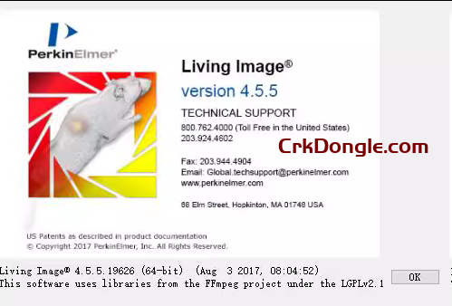
Overview of Living Image 4.5
Living Image is a powerful image acquisition and analysis software developed by PerkinElmer (now Revvity) for their IVIS® in vivo imaging systems. It supports preclinical research in oncology, infectious diseases, drug development, and neuroscience by enabling 2D and 3D visualization, quantification, and analysis of bioluminescent and fluorescent signals in live animals. The software processes data from optical, CT, and multi-modality imaging, offering tools for ROI drawing, spectral unmixing, tomographic reconstruction (DLIT and FLIT), and co-registration with anatomical images. It integrates with IVIS Spectrum, Lumina, and other systems, providing workflow automation, customizable reporting, and export options to formats like TIFF, AVI, and Excel. Living Image is widely used in academic and pharmaceutical labs for non-invasive, longitudinal studies.
Key Features of Living Image
- Image Acquisition: Real-time control of IVIS hardware for bioluminescence, fluorescence, and CT imaging; supports sequence acquisition with automated focus and temperature control.
- 2D Analysis: ROI tools for signal quantification, spectral imaging for separating overlapping fluorophores, and background subtraction.
- 3D Tomographic Reconstruction: DLIT (Diffuse Light Imaging Tomography) for bioluminescence depth and location; FLIT (Fluorescence Lifetime Imaging Tomography) for lifetime-based analysis; one-click reconstructions and multi-subject overlays.
- Multi-Modality Integration: Co-registration of optical data with CT for anatomical context; 3D volume rendering and surface mapping.
- Visualization and Reporting: Sequence views for time-series analysis, 3D ROI synchronization across views, photo-realistic rendering, and customizable dashboards.
- Advanced Modules:
- 3D Multi-Modality Module: For enhanced CT-optical fusion and volumetric analysis.
- Sequence Builder: Automates multi-step imaging protocols.
- Spectral Unmixing: Algorithms for precise fluorophore separation in multiplexed studies.

